Beyond text
- Nurses
- Students
- Educators
Images
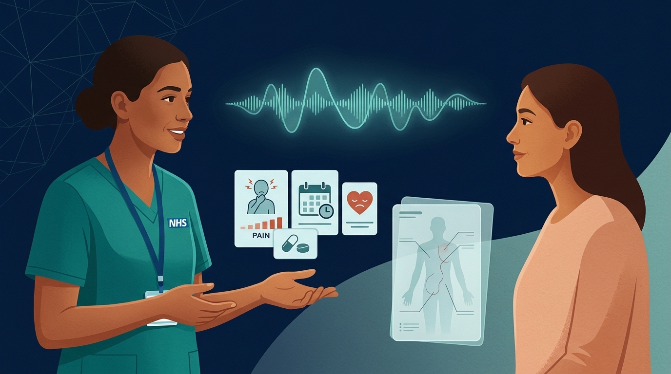
Never upload patient photos, wounds, rashes, or charts into a general AI tool — even with faces cropped out.
AI image models default to light skin tones — pair educational visuals with resources like Mind the Gap.
Verify anatomy, device placement, and embedded labels with a qualified clinician before teaching or sharing.
Ways images and AI meet in nursing
Diagrams & teaching visuals
Creating conceptual illustrations and visual analogies for teaching and patient education (always verified by a clinician).
Drafting alt text
Writing screen-reader descriptions for non-confidential teaching slides — edit to explain why the image matters clinically.
Never upload patient images
Never upload photographs of patients, wounds, rashes, charts or screens into a general AI tool, even with faces hidden.
Never upload identifiable images
Don't put photographs of patients, wounds, skin, documents, screens or anything else identifiable into a general AI tool, even with the face hidden. Wound and skin images are personal health data. Analysing them is a clinical activity that needs an approved, clinically assured system. Some image analysis tools are regulated medical devices.
Where image generation helps
- Explaining how things work: how an inhaler reaches the lungs, how a stoma works, where a line sits. See Medication mechanisms.
- Teaching: concept diagrams, visual mnemonics and scenario illustrations.
- Accessible information: simple, consistent pictures to support easy read. These need checking with people who use easy read. See Accessible information.
Where it can cause harm
| Risk | What to check |
|---|---|
| Anatomical and clinical errors | Extra fingers and impossible anatomy are obvious. Wrong placement of a device, wrong wound stages or wrong technique are not. Check every clinical image with someone qualified. |
| Who is shown and how | AI image tools often default to particular skin tones, body types, ages and genders. Clinical signs such as rashes, cyanosis and pressure damage look different on different skin, and teaching images have historically under-represented Black and brown skin (Mind the Gap). Check your images show the range of people you care for. |
| Misleading realism | Photorealistic images can look like real patients or real evidence. Label generated images as illustrations. |
| Text in images | AI-generated text in images is often misspelt or wrong. Check every word, or add text yourself. |
| Copyright and likeness | Don't ask for images in the style of a living artist, or that resemble real people. |
Good practice for alt text
AI can draft alt text quickly, but it describes what it sees, not what the image is for. Edit it to say why the image matters in this context. For example, write "Diagram showing the inhaler spacer attached to the mouthpiece", not "A grey object on a white background".
Try it
Create a simple, clearly labelled illustration for patient education showing
how a spacer device attaches to an inhaler. Plain style, no text other than the labels
"inhaler", "spacer" and "mouthpiece". Then write alt text explaining what the image shows and why.
Check the result against the manufacturer's instructions before using it with anyone.
A colleague crops a patient's face out of a smartphone photo of a sacral pressure ulcer and asks if they can upload it to a general AI chatbot to help grade the wound category. What is the correct response?